
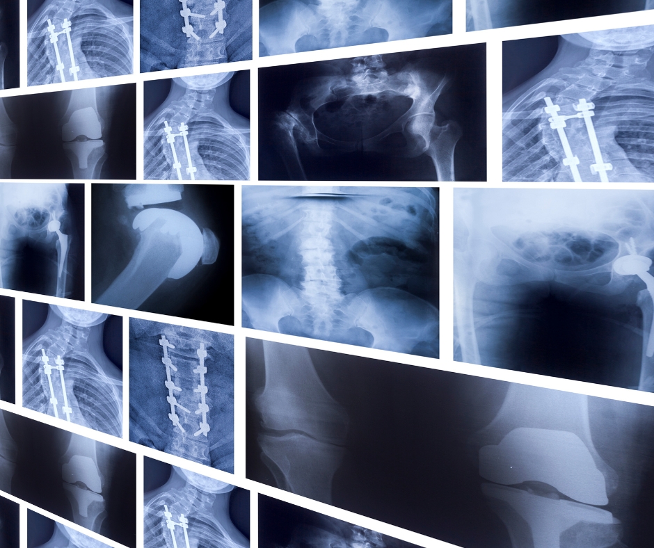
Understanding the difference between a CT scan and a standard X-ray helps patients feel more informed and prepared when referred for medical imaging. While both diagnostic techniques rely on X-ray radiation to create images of the inside of the body, they serve distinctly different clinical purposes. Choosing between them depends on the specific organ, structure, or condition your doctor needs to evaluate.
The Difference Between Two-Dimensional and Three-Dimensional Imaging
The fundamental difference between an X-ray and a CT scan lies in how the image is captured and rendered. A standard X-ray passes a single beam of low-dose radiation through the body onto a digital detector plate behind the patient. This process creates a flat, two-dimensional image similar to a shadow photograph. Denser structures like bones absorb more radiation and appear bright white, while softer tissues like lungs allow radiation to pass through and appear dark.
A computed tomography (CT) scan takes this process further by combining multiple X-ray measurements into detailed cross-sectional views. During a CT scan, an X-ray source rotates around the patient inside a circular housing, acquiring hundreds of thin digital slices. A computer processes these slices to construct three-dimensional views of internal organs, soft tissues, blood vessels, and complex bony structures.
Why Medical Imaging Choices Matter
Selecting the appropriate imaging modality directly influences diagnostic accuracy, treatment planning, and patient safety. Standard X-rays are exceptionally efficient for quick, first-line evaluations. They allow radiographers to assess acute fractures, joint dislocations, dental issues, and basic chest conditions in a matter of seconds.
However, two-dimensional X-rays have clear limitations due to tissue overlapping. Structures in the foreground can obscure soft tissues or subtle abnormalities located directly behind them. CT scanning resolves this overlap by isolating thin slices of anatomical structure. This detailed clarity allows radiologists to detect tiny lung nodules, complex bone fractures, internal bleeding, abdominal inflammation, and vascular disease that remain invisible on standard X-ray films.
Practical Advice for Patients
If your referring medical practitioner orders an imaging procedure, understanding these practical differences can help you prepare for your appointment:
- Procedure Duration: A standard X-ray takes only a few minutes to set up and complete. A CT scan requires slightly more time, typically lasting between 10 and 20 minutes for patient positioning, scanner preparation, and image acquisition.
- Radiation Exposure: CT scans gather vastly more detailed data than flat X-rays, which means they involve a higher dose of ionising radiation. Radiographers always follow strict protocol guidelines to keep radiation exposure as low as reasonably achievable while maintaining diagnostic quality.
- Contrast Agents: Standard X-rays rarely require contrast materials. CT scans frequently utilise oral or intravenous contrast dye to highlight blood vessels, digestive tracts, and soft tissue structures.
Expert Recommendations for Choosing Imaging Modalities
Medical imaging guidelines recommend starting with the least invasive and lowest radiation option that can reliably answer the clinical question. Standard X-rays remain the preferred initial tool for routine orthopedic checks, immediate trauma triage, and general chest screening.
When clinical symptoms suggest complex internal conditions, your general practitioner or specialist will refer you for a CT scan. CT is the gold-standard modality for investigating acute abdominal pain, evaluating stroke symptoms, staging oncological conditions, and planning complex surgical interventions. Always discuss any previous imaging history or potential pregnancy with your imaging clinic before undergoing a scan.
Common Mistakes in Medical Imaging Expectations
A frequent misconception among patients is assuming that a CT scan is always superior to a standard X-ray for every health issue. While CT offers higher detail, an X-ray is often safer, faster, and completely sufficient for straightforward conditions like a suspected wrist fracture or basic chest clearings. Expecting a CT scan for simple injuries unnecessarily increases radiation exposure and medical costs.
Another common mistake is confusing CT scans with Magnetic Resonance Imaging (MRI). While CT uses X-rays to image high-density structures and emergency conditions, MRI uses strong magnetic fields and radio waves to capture highly detailed images of soft tissues, ligament tears, and neurological structures without using ionising radiation.
Summary
Standard X-rays provide fast, two-dimensional images that are ideal for basic bone and chest evaluations. CT scans rotate X-ray equipment around the patient to generate highly detailed three-dimensional cross-sections of complex soft tissues, organs, and blood vessels. Both procedures play vital roles in modern medicine, and your referring doctor will select the safest, most effective imaging tool for your unique diagnostic requirements.
Key Takeaways
- Standard X-rays produce flat two-dimensional images, while CT scans create three-dimensional cross-sectional slices.
- X-rays are best suited for initial assessments of bone fractures, joint health, and basic lung conditions.
- CT scans are essential for examining complex soft tissue, internal organs, blood vessels, and hidden trauma.
- Radiation exposure is higher in CT scanning due to the multiple images required to construct three-dimensional views.
- Your medical doctor determines the correct test based on diagnostic necessity, speed, and safety considerations.